Upper Thigh Muscle Anatomy / Involved early gray = muscle:. Horizontal groove on the posterior aspect of medial condyle of. Leg muscle anatomy for figurative artists. Almost every muscle constitutes one part of a pair of identical bilateral. We think this is the most useful anatomy picture that you need. Anatomy of the human body.
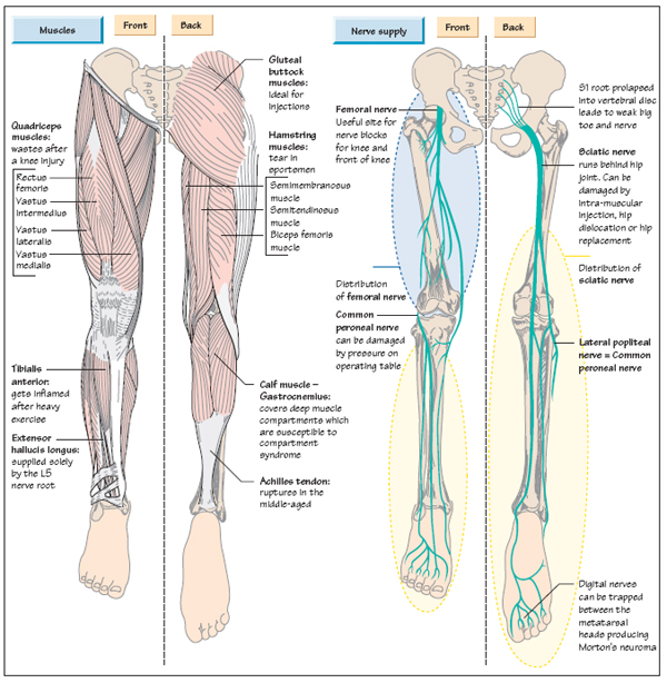
Because of their broad attachments this muscle contributes to most of the flesh of the buttocks. The muscles and fasciæ of the thigh. The sartorius muscle attaches to the hip bone (iliac spine), travels down the front of the thigh moving toward the inside of the thigh, and connects to the inside of the shin bone (tibia). A complete list of muscular system quizzes; Musculoskeletal anatomy, kinesiology, and palpation for manual therapists.
Anatomy of the Leg | Musculoskeletal Key from musculoskeletalkey.com The trapezius muscles are superficial muscles of the neck and upper trunk. 2, vastus medialis & intermedius muscles. It is part of the lower limb. The muscle passes out of the pelvis through the greater sciatic foramen, the upper part of which it fills, and is inserted by a rounded tendon into the upper border of the greater trochanter behind, but often partly blended with. Thigh muscle anatomy hip anatomy gross anatomy yoga anatomy human body anatomy human anatomy and physiology anatomy study anatomy reference leg muscles anatomy. This is a table of skeletal muscles of the human anatomy. Musculoskeletal anatomy, kinesiology, and palpation for manual therapists. Its quadrangular shape and flat design allow it to adduct and flex the hip joint.
Leg muscle anatomy for figurative artists.
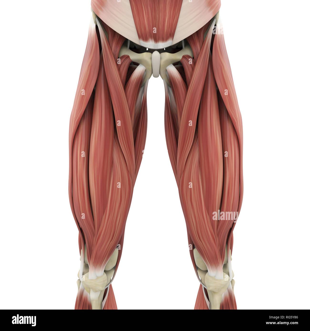
The muscles and fasciæ of the thigh. Involved early gray = muscle: The single bone in the thigh region is called the femur. The uppermost of the medial thigh muscles is the pectineus muscle. You can click the links in the image, or the links below the image to find out more information on any muscle group. Superolateral part of upper quadtrilateral area of ischial tuberosity. Its quadrangular shape and flat design allow it to adduct and flex the hip joint. The pectineus is a flat, quadrangular muscle situated at the anterior part of the upper and medial aspect of the thigh. The sartorius muscle attaches to the hip bone (iliac spine), travels down the front of the thigh moving toward the inside of the thigh, and connects to the inside of the shin bone (tibia). We think this is the most useful anatomy picture that you need. It is part of the lower limb. This image added by admin. Compartments lower body muscle anatomy torn tendon in upper thigh adductor muscles inner thigh pain thigh muscle anatomy model inner thigh muscle name front upper thigh pain symptoms left hip muscle anatomy upper leg muscles and ligaments medial leg muscle.
This is a table of skeletal muscles of the human anatomy. This muscle moves the upper leg. The thigh is the area between the hip and th. Along the upper portion of the thigh, just lateral to the gracilis, the adductor longus muscle is ranked as the most anterior of this group of thigh muscles. You can click the image to magnify if you cannot see clearly.
Upper Body Skeleton High Resolution Stock Photography and ... from c8.alamy.com The hamstring muscles include (all the muscles of posterior compartment of thigh except short head of biceps femoris): Find the best weight lifting exercises that target each muscle or groups of muscles. The muscular effort (contraction) is applied to the bone at the insertion of the muscle and produces motion if the effort exceeds the resistance (load). The thigh is the area between the hip and th. The muscles and fasciæ of the thigh. Involved early gray = muscle: The uppermost of the medial thigh muscles is the pectineus muscle. This muscle moves the upper leg.
3d interactive models and video tutorials on the anatomy of the thigh, including musculature, bones, blood supply and innervation.
Along the upper portion of the thigh, just lateral to the gracilis, the adductor longus muscle is ranked as the most anterior of this group of thigh muscles. The muscle passes out of the pelvis through the greater sciatic foramen, the upper part of which it fills, and is inserted by a rounded tendon into the upper border of the greater trochanter behind, but often partly blended with. Thigh muscle anatomy hip anatomy gross anatomy yoga anatomy human body anatomy human anatomy and physiology anatomy study anatomy reference leg muscles anatomy. Let's begin with the skeletal anatomy. The pectineus is a flat, quadrangular muscle situated at the anterior part of the upper and medial aspect of the thigh. There are around 650 skeletal muscles within the typical human body. 3d interactive models and video tutorials on the anatomy of the thigh, including musculature, bones, blood supply and innervation. Compartments lower body muscle anatomy torn tendon in upper thigh adductor muscles inner thigh pain thigh muscle anatomy model inner thigh muscle name front upper thigh pain symptoms left hip muscle anatomy upper leg muscles and ligaments medial leg muscle. This muscle moves the upper leg. This webpage presents the anatomical structures found on thigh mri. Leg muscle anatomy for figurative artists. The thigh is the area between the hip and the knee joint. The single bone in the thigh region is called the femur.
Muscles and ligaments work together to support the spine, hold it upright, and control movement during rest and activity. Superolateral part of upper quadtrilateral area of ischial tuberosity. The muscular effort (contraction) is applied to the bone at the insertion of the muscle and produces motion if the effort exceeds the resistance (load). The uppermost of the medial thigh muscles is the pectineus muscle. It is part of the lower limb.
Muscles of the Thigh and Gluteal Region - Part 2 - Anatomy ... from i1.ytimg.com Muscles are named according to their shape, location, or a combination. Find the best weight lifting exercises that target each muscle or groups of muscles. 1.1 how skeletal muscles produce movement. Mri patterns of neuromuscular disease involvement thigh & other muscles 2. It is used primarily when the hip is already flexed. This webpage presents the anatomical structures found on thigh mri. We think this is the most useful anatomy picture that you need. Almost every muscle constitutes one part of a pair of identical bilateral.
This muscle moves the upper leg.
The thigh is the area between the hip and th. Superolateral part of upper quadtrilateral area of ischial tuberosity. You can click the image to magnify if you cannot see clearly. Thigh muscle anatomy hip anatomy human body anatomy yoga anatomy human anatomy and physiology anatomy study anatomy reference leg muscles anatomy pose reference. Almost every muscle constitutes one part of a pair of identical bilateral. Basic anatomy terminology | kenhub anatomy guide. Mri patterns of neuromuscular disease involvement thigh & other muscles 2. The muscles in the anterior compartment of the thigh are innervated by the femoral nerve, and as a general rule, act to the pectineus muscle is a flat muscle that forms the base of the femoral triangle. Leg muscle anatomy for figurative artists. Find the best weight lifting exercises that target each muscle or groups of muscles. The sartorius muscle attaches to the hip bone (iliac spine), travels down the front of the thigh moving toward the inside of the thigh, and connects to the inside of the shin bone (tibia). Anatomynote.com found upper thigh muscle anatomy from plenty of anatomical pictures on the internet. The muscular effort (contraction) is applied to the bone at the insertion of the muscle and produces motion if the effort exceeds the resistance (load).
2, vastus medialis & intermedius muscles upper thigh anatomy. 1.1 how skeletal muscles produce movement.


Posting Komentar
0 Komentar